Tuesday 30 November 2010

Bringing order to the Virosphere


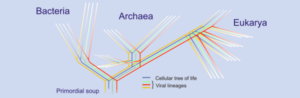
Putative tree of life including viral sequences. The 'sea' of viruses. http://blogs.helsinki.fi/bamford-group/files/2008/06/vevo.png

Physically and evolutionarily speaking we are bathed in a sea of viruses and virus-like elements. Over the last billion or so years these genetic parasites have affected the lives of all kinds of organisms worldwide with sometimes devastating consequences (and others somewhat more positive). An extremely conservative estimate of virus numbers is more likely in the billions and so a major issue is just exactly how can we make sense of all this diversity out there in the Virosphere


Determining how one virus – or group of viruses – is related to any other is pretty straightforward nowadays; with sequencing technology and bioinformatic analysis commonplace, we can effortlessly compare nucleotide sequences and generate phylogenetic trees displaying how two or more relate to each other.  This works pretty well in a modern setting when we can compare viral isolates from patients infected in a certain outbreak and determine the path of spread for example over days, weeks, months or years,


Phylogenetic tree tattoo. http://blogs.discovermagazine.com/loom/files/2008/07/origin.jpg

This kind of analysis may however have its limits when we attempt to apply it to virus groups that have potentially diverged millions of years ago – a scenario which would be expected given that viruses have been around with us since the beginning of life itself. Simply over that length of time, any evolutionary ‘signal’ found within a particular gene sequence will have disappeared. As masters of genetic variation, virus genomes can get pretty messy, evolutionary speaking due to horizontal gene transfer, recombination and high mutation rates. How can we then assess the deeper evolutionary questions with regards these highly variable agents?


Krupovic and Bamford (who both specialise in the origins and early evolution of viruses), commenting in the Journal of Virology recently, suggest that we should be focussing our analysis at a different aspect of viral lifestyles: the structural biology of their virions – a feature which they believe reflects the true nature of what a virus really is. This, they say should be suggestive of the early and highly conserved evolutionary relationships (nucleotide sequence may be variable but protein sequence/structure will be more highly conserved). By adopting this strategy, the problems of horizontal gene transfer may also be ablated making all variation observed relative to this gene/protein architecture. Using this perspective, we could infer higher taxonomic structures for the Virosphere reflecting true, ancient biological relationships – that seemingly unrelated viruses infecting divergent host species share a common origin (which has been seen for some human and bacterial viruses).


Picornavirus virion structure made up of viral capsid proteins.  Illustration by David S. Goodsell of The Scripps Research Institute (see this site) [Public domain], via Wikimedia Commons


By attempting to group distantly related viruses, maybe those infecting bacteria, archaea and eukaryotes into common taxonomic classes we should be able to bring more order and structure to the viral universe. – facilitating a better understanding of the entire tree of life. We will not, however be able to accomplish this unless we realise the true age of these viruses and adapt our evolutionary investigations for this. Caveats aside, the idea presented by Krupovic and Bamford should stimulate further evolutionary work to be carried out on more virus taxa.

Order to the Viral Universe. Krupovic and Bamford J. Virol..2010; 84: 12476-12479

Sunday 21 November 2010

Epithelial defence – the role of highly reactive oxygen species

Cell-cell communication is a key feature of biological systems particularly multicellular eukaryotes whose development and functioning rely on complex information exchange between and within cells and tissues and across both long and short distances. The relative roles of particular cell-cell signalling pathways is therefore of interest to a number of processes - one being the host responses during infection.

Fig.1. Typical epithelial cell layer (lung tissue in this case). Nuclei stained in blue, actin in red and tight junctions in green. Notice the physical barrier formation. http://www.seas.upenn.edu/~injury/images/091201%20triple%20slide%20test%20A5%201_5para%2040x%20overlay.jpg


Friday 19 November 2010

Stopping pathogens at the portal of entry: mucous membrane defence


Enhancing Oral Vaccine Potency by Targeting Intestinal M Cells.
Azizi A, Kumar A, Diaz-Mitoma F, Mestecky J (2010)

The immune system in the gastrointestinal tract plays a crucial role in the control of infection, as it constitutes the first line of defense against mucosal pathogens. The attractive features of oral immunization have led to the exploration of a variety of oral delivery systems. However, none of these oral delivery systems have been applied to existing commercial vaccines. To overcome this, a new generation of oral vaccine delivery systems that target antigens to gut-associated lymphoid tissue is required. One promising approach is to exploit the potential of microfold (M) cells by mimicking the entry of pathogens into these cells. Targeting specific receptors on the apical surface of M cells might enhance the entry of antigens, initiating the immune response and consequently leading to protection against mucosal pathogens. In this article, we briefly review the challenges associated with current oral vaccine delivery systems and discuss strategies that might potentially target mouse and human intestinal M cells.


Different parts of our bodies are not equally vulnerable to infection and we can consider our immune system to be differentially active across all anatomical sites. One of the biggest sites of pathogen entry is the mucosal epithelium which lines our internal parts including the respiratory, gastrointestinal and urogenital tracts (fig 1.).

These surfaces have been estimated to cover 200x the area that our skin covers and if you think about it, most pathogens will use one of these openings to the outside world to gain entry into our bodies: measles virus via the respiratory tract; HIV through the urogenital tract; and E.coli from the gastrointestinal tract. It is this reason that we expend so much energy and effort in keeping these areas protected from would-be dangerous micro-organisms.

Fig. 2. Dipiction of the mucosal immune system:inductive and effector sites both present with mechanisms of inductive and nuetralisation (SIgA) shown. Notice the MALT complex (M cells, antigen presenting cells and B/T cells). http://www.nature.com/mi/journal/v1/n1/fig_tab/mi20079f1.html


Mucosal immunity is a complex system of anatomical, cellular and molecular 'innate' components: epithelial cells which line the mucosa form very tight layers which prevent bacteria or viruses from penetrating deeper into the body; these cells also secrete a number of antimicrobial compounds and enzymes and can rapidly respond to infection through protein mediated cell-cell signalling establishing intra-cellular protection.

In order to mount an efficient response there is a close association between the mucosal epithelium and lymphoid tissues. This 'mucosa-associated lymphoid tissue' or MALT (examples including nasal-associated lymphoid tissue or NALT and gut-associated ymphoid tissue or GALT) is formed by a close interaction between the epithelial lining (antigen sampling 'M' cells) and underlying immune cell complexes consisting of antigen-presenting cells, T cells and B cells.

These mucosal sites – interestingly – form a collective adaptive unit across the whole body when we consider anti-microbial immunity. This 'common mucosal immune system' is mediated by a network of inductive and effecter sites located within the MALT: inductive sites sensing pathogen activity at one area can generate cellular immune responses which can travel systemically from one site to another (e.g. NALT to GALT) . One of the major players in this immunity is a special type of antibody known as secretory immunoglobulin A (SIgA) which is transported from the underlying immune tissue at effector sites to the surface of the mucosa above and can interact with and bind to pathogens and prevent infection and/or damage being done (see fig2 and fig3).

Interest has stemmed from the thought that if we were able to generate adequate antibody responses at the pathogen site of entry (SIgA at the mucosa) we may able to prevent infection from occurring before it is too late and infection has spread from primary areas to other tissues causing significant disease . Current vaccination strategies have focussed on the development of systemic IgG antibodies without consideration for those found at initial portals of entry and although many have been successful for some diseases, the millions of deaths stemming from pathogen-mucosa related infections warrant further investigation and targeting. The question is which inductive MALT site is best suited for vaccination which in turn depends upon pathogen entry strategy, ease of administration, particular side effects and the relative ability of particular MALTs to develop effecter functions at different mucosal epithelium. To date the most promising of strategies lies with nasal or oral vaccine delivery systems and much research has been carried out in order to improve the efficacy of each.

Fig 3. Cartoon dipicting the general structure of the five basic antibodies in humans: IgA the one concerned with mucosal immunity. http://www.cartage.org.lb/en/themes/sciences/lifescience/generalbiology/physiology/LymphaticSystem/Antibodymediated/AntiBtypes.gif

Novel approaches have been used to target vaccines to those cells responsible for antigenic sampling found across most mucosal surfaces – the M cells. These cells can transport antigenic materials from the epithelial lining to the underlying lymphoid complexes via antigen-presenting cells. These cells however are present in low numbers (1 in 10 million epithelial cells are M cells in the GI tract) with the mucosa hence specific targeting may facilitate higher antigen uptake and greater immune induction and these strategies can be adapted to both respiratory and oral delivery systems depending on the vaccine.

We are only now beginning to see the outcome of current research into mucosal immunity and vaccines. Disrupting the close relationship between pathogens and the mucosa is paramount in preventing the millions of deaths worldwide from mucosal-borne infections and the concurrent development of novel targeting and vaccination strategies we may well witness the generation of highly effective, easily administered and most importantly safe vaccines for the most dangerous known diseases: AIDS; cholera; and influenza.

Azizi A, Kumar A, Diaz-Mitoma F, Mestecky J (2010) Enhancing Oral Vaccine Potency by Targeting Intestinal M Cells. PLoS Pathog 6(11): e1001147. doi:10.1371/journal.ppat.100114


Thursday 11 November 2010

Pathogenesis and cell-cell spread of a deadly virus

Fig.1 NiV infection in an epithelial cell monolayer and different time points and stained (red). Epithelial adhesion indicated in green.

Nipah virus (NiV), an emerging and deadly paramyxovirus of bat origin has been responsible for a number of fatal outbreaks of disease in humans and other animals in South-East Asia and as of yet no vaccine or therapy exists. The mucosal epithelium of the host respiratory tract is believed to be the most common route of viral spread in humans and from this primary site the virus can disseminate systemically through the blood system resulting in rapid endothelial dysfunction and vasculitis. Viruses must overcome a number of cellular mechanical barriers in order to establish efficient infection and knowledge of how they do this is key to understanding pathogenesis and infection in general.

The ability to move between and within cells is central to viral transmission and pathogenesis. Viruses - as obligate intracellular parasites – need to effectively enter and exit target cells within the host organism in which they reside and establish productive infections to facilitate transmission and replication. Viruses generally use two methods of spreading – cell free and cell associated transmission; the first requiring assembly and budding of mature virus from the cell membranes and the second taking advantage of the close association between host cells, facilitating the union of the membranes of two separate cells. The relative roles of each of these processes in vivo and in transmission and infection are not fully known.

The NiV F and G glycoproteins are responsible for virion-host cell membrane fusion and also cell-cell fusion via interactions with NiV receptors expressed in the plasma membrane of target cells. The mechanism of viral spread within the host are thus very important in elucidating how the virus causes disease and transmits itself within a population; polarised epithelial surfaces – where the apical and basolateral domains are structurally and functionally distinct–playing dual roles in initial entry and host-host viral spread.

Fig.2. NiV virion - notice the fusion and attachement proteins on the surface (red and green). http://www.bepast.org/docs/photos/Nipah%20Virus/nipah%20virus.jpg.

Viruses have taken advantage of the fact that host cells require discrete functions on their apical and basolateral domains and can preferentially target mature virus formation to particular cellular locations through intrinsic protein signals built in to their glycoproteins. Apical release will generally facilitate free virion spread and host-host transmission (e.g in the respiratory tract) while basolateral release may allow direct cell-cell spread and could lead to systemic infections not restricted to the particular primary site of replication. NiV can therefore control its transmission within and between hosts by targeting its infection machinery (F and G glycoproteins) to either the apical or basolateral surfaces of polarised cells.

Fig.3. Polarised epithelial cell monolayer. Apical and basal domains shown. http://www-dsv.cea.fr/var/plain/storage/original/media/File/IBITECS/SB2SM/Eq%20Verbavarz/Cell-base.JPG

A recent paper has investigated the mechanism of NiV spread in polarised epithelium in vitro using cell imaging and molecular analyses of its F and G glycoproteins. They demonstrate that the NiV glycoproteins can facilitate cell-cell spread within polarised epithelial cells through targeting to basolateral cell surfaces between closely positioned cells allowing entry to epithelial cells or underlying tissues. They also note that they are also found on the apical domain allowing efficient spread via free virus release. The group explored the functional importance of such targeting by determining the exact molecular signals required for such protein trafficking and then disrupting them. By purposely retargeting the fusion machinery to the apical side, NiV cell-cell spread by lost. The group also report their findings of NiV infection within infected endothelial cells – a major target of NiV – and establish that this interaction may allow viral entry into the central nervous system (CNS).

Their results add to the ongoing the investigation of not only NiV pathogenesis but also to that of other important and deadly viral pathogens. The initial site of viral entry and its subsequent release and spread within a host are essential to the degree of infection and pathogenesis caused by a particular virus in its host. Spread of NiV into sub-epithelial tissues facilitates systemic infection through the host vasculature and leads to the establishment of unique sites of replication in organs systems and tissues far away from the initial portal of entry. Apical dissemination of infectious virus will also allow for host-host transmission via respiratory or urogenital epithelia.

Knowledge of how the virus achieves this spread may allow us to design better vaccines, identify novel therapeutic targets and add to the wealth of information in viral pathogenesis.


Carolin Weise, Stephanie Erbar, Boris Lamp, Carola Vogt, Sandra Diederich, and Andrea Maisner. Tyrosine Residues in the Cytoplasmic Domains Affect Sorting and Fusion Activity of the Nipah Virus Glycoproteins in Polarized Epithelial Cells. Journal of Virology, August 2010, p. 7634-7641, Vol. 84, No. 15

Friday 29 October 2010

Viral genomes in High Definition

As seen with cellular organisms, our ability to rapidly and accurately sequence multiple viral genomes at low cost is being overtaken by our inability to analyse the functional roles that certain genes – including regions within genes- play in a range of phenotypic processes during an infection. Lack of functional studies represents a major roadblock to understanding the basic biology of these pathogenic organisms and prevents us from generating safe and efficacious vaccines or virotherapies.

High-Resolution Functional Mapping of the Venezuelan Equine Encephalitis Virus Genome by Insertional Mutagenesis and Massively Parallel Sequencing

Brett F. Beitzel, Russell R. Bakken, Jeffrey M. Smith, Connie S. Schmaljohn*

The United States Army Medical Research Institute of Infectious Diseases, Fort Detrick, Maryland, United States of America

We have developed a high-resolution genomic mapping technique that combines transposon-mediated insertional mutagenesis with either capillary electrophoresis or massively parallel sequencing to identify functionally important regions of the Venezuelan equine encephalitis virus (VEEV) genome. We initially used a capillary electrophoresis method to gain insight into the role of the VEEV nonstructural protein 3 (nsP3) in viral replication. We identified several regions in nsP3 that are intolerant to small (15 bp) insertions, and thus are presumably functionally important. We also identified nine separate regions in nsP3 that will tolerate small insertions at low temperatures (30°C), but not at higher temperatures (37°C, and 40°C). Because we found this method to be extremely effective at identifying temperature sensitive (ts) mutations, but limited by capillary electrophoresis capacity, we replaced the capillary electrophoresis with massively parallel sequencing and used the improved method to generate a functional map of the entire VEEV genome. We identified several hundred potential ts mutations throughout the genome and we validated several of the mutations in nsP2, nsP3, E3, E2, E1 and capsid using single-cycle growth curve experiments with virus generated through reverse genetics. We further demonstrated that two of the nsP3 ts mutants were attenuated for virulence in mice but could elicit protective immunity against challenge with wild-type VEEV. The recombinant ts mutants will be valuable tools for further studies of VEEV replication and virulence. Moreover, the method that we developed is applicable for generating such tools for any virus with a robust reverse genetics system.


Beitzel et al recently publish the development of a ‘high-resolution genomic mapping technique’ that will facilitate the easier investigation of particular viral genes in a range of virus/host interactions. Venezuelan equine encephalitis virus, endemic to South America, is an important zoonotic pathogen that continually re-emerges from its rodent reservoir via a mosquito vector causing potentially fatal encephalitis in both humans and horses. There is currently no vaccines or anti-viral treatments for this disease and the exact roles that each of its 8 proteins play in its basic biology and replication are currently being investigated, hoping to yield helpful insight.

Studying the role of nsP3 gene in VEEV but later expanding it the rest of the genome, the group used a technique called transposon mutagenesis in which a short (15 nucleotides) DNA fragment is inserted randomly into the target gene or genome being studied. They generated a large cDNA library with high coverage in that every nucleotide had been targeted around 200 times and using high- throughput DNA sequencing were able to map the inserts at a high resolution and record the frequency that they were present at. Combining this with a reverse genetics system, they generated recombinant infectious virus carrying and expressing the inserts in order to assess their function in replication and initially, the group assessed the role that these inserts had on replication at particular temperatures.

By altering the temperatures in which the viruses were grown they were to able to effectively screen for a particular phenotype (the ability to grow at 40 degrees for example) and easily map the locations that would and would-not tolerate the 15bp insertions. Using this method they generated temperature sensitive (ts) mutants that were able to replicate at one temperature (30) and not at another (40).


Monday 18 October 2010

Viral control with endosymbiotic bacteria - the dengue story

No sooner did we hear that we have effectively caused the extinction of one important virus from the earth, we get news of the dramatically increasing incidence of mosquito-borne Dengue virus (DENV) infection worldwide with the numbers doubling over the last decade (two-fifths of the global population now at-risk estimated at 2.5 billion individuals). Dengue is a textbook example of an emerging disease although has been largely ignored in most developed countries as it is mostly found in urban districts of tropical/sub-tropical countries. It is now considered the most important arthropod-borne viral disease and developed countries should take note as DENV is predicted to expand its range with a changing climate.

Fig.1. WHO stats on DENV distribution 2005

Dengue viruses cause what is known as a severe “flu-like” illness including symptoms such as rash, mild/high fever, headache and muscle pain. Although it is rarely fatal, in some instances it can develop into Dengue Haemorrhagic Fever (DHF), a potentially deadly complication. There are generally considered four sub-types (DENV-1 to 4) of Dengue virus due to the sufficient antigenic differences exhibited between them leading to little or no effective cross-neutralisation making it difficult to develop a ‘universal’ Dengue vaccine or treatment. The viruses are spread by certain Aedes species of mosquito passing infectious virus on during feeding by the females and due to the lack of treatments, the inhibition of mosquito activity has been seen as key to Dengue prevention.

A number of ‘vector control’ methods are being carried out to limit mosquito-human viral transmission which focus on disrupting the ecological niche in which the vector requires to breed – in this case stagnant water pools found in urban areas in which larva are found (e.g. rainwater filled tyres and cups). One major strategy is the biological control of Aedes populations including the use of mosquito larvae preying fish and invertebrates, release of Aedes infecting viruses or by genetic modification of mosquito genomes. An attractive means of control has come from the realisation that certain bacterial species (Wolbachia) - and only a specific strain of it - which naturally infects mosquitoes and other insects could inhibit dengue virus transmission thus negating the need for transgenic mosquitoes in the environment. These obligate, intracellular parasites which survive in the host cell’s cytoplasm are passed maternally from generation to generation. The bacteria can affect the mosquitoes and dengue virus in a number of ways, both general and specific for example: reduced lifespan of Aedes hosts; reduced dengue virus replication; increased antiviral immunity; and spatial exclusion of virus from the cytoplasm.

Fig.2. DENV transmission in Aedes species http://activity.ntsec.gov.tw/lifeworld/english/content/images/en_dis_c10.jpg

Frentiu et al report their investigations into the mechanism of reduced viral (dengue virus serogroup 2) replication at the cellular level paralleling earlier observations of whole organism Wolbachia infection. Their transition to a cell-culture experimental system may facilitate easier study of viral-host interactions and the group documents significantly reduced dengue virus replication in Wolbachia infected cell lines compared to non-infected controls possibly being related to an increased bacterial density within the cytoplasm. A bacterial ‘priming’ of the insect immune system may also contribute to decreased replication. They also show evidence that Wolbachia infected mosquitoes may display a fitness benefit compared to those not infected when challenged with dengue virus. This predicted increased fitness in the wild may aid the use of this biological control technique in a natural ecosystem. Measures such as these will benefit from the large, field trials already planned to study Aedes-Wolbachia interactions.

Fig.3. Wolbachia (Green) within a drosophila embryo imaged by confocal microscopy. http://www.genetics.org/content/vol178/issue4/cover.dtl

Dengue virus is an important emerging arthropod-borne pathogen worldwide and is predicted to further increase its range into more temperate regions. Currently there is no effective vaccine or treatment and much research has focused on the interactions between virus and Aedes mosquito including the use of intracellular bacterial pathogens to limit viral replication and transmission. A number of strategies have been studied and seem attractive on a wider scale in endemic countries however the mechanisms of DENV/Wolbachia interactions and the effects on arthropods in the wild are understudied. Frentiu et al highlight the importance of a mechanistic understanding of dengue control and the development of novel control strategies.

Wednesday 13 October 2010

Of owls and viruses

Viral Research in Brazilian Owls (Tyto alba and Rhinoptynx clamator) by Transmission Electron Microscopy

*Catroxo, M. H. B.; *Taniguchi, D. L.; *Melo, N. A.; **Milanelo, L.; ***Petrella, S.; **Alves, M.; *Martins, A. M. C. R. P. F. & *Rebouças, M. M.


SUMMARY: The barn-owl (Tyto Alba) and striped-owl (Rhinoptynx clamator) belong respectively to the families Tytonidae and Strigidae. Avian paramyxoviruses have been isolated from a variety of species of wild and domestic birds wordlwide causing diverse clinical symptoms and signs. Paramyxoviruses belong to the family Paramyxoviridae and Avulovirus genus, including nine serotypes (APMV 1 to 9). The lymphoid leukosis is a retrovirus-induced neoplasia. The avian retroviruses belong to the Retroviridae family and to the Alpharetrovirus genus. Coronaviruses can cause respiratory and enteric disease in several species of birds. They belong to the Coronaviridae family and to the groups 3a e 3c. In this study, we describe the presence of viruses in four owls, two barn owls (Tyto alba) and two striped owls (Rhinoptynx clamator), rescued from tree-lined streets of Sao Paulo, Brazil and sent to the Recovery Center of Wild Animals of the Tietê Ecological Park, where the animals died. Fragments of lung, liver and small intestine of these birds were processed for transmission electron microscopy utilizing negative staining (rapid preparation), immunoelectron microscopy and immunocitochemistry techniques. Under the transmission electron microscopy paramyxovirus particles, pleomorphic, roughly spherical or filamentous, measuring 100 to 500 nm of diameter containing an envelope covered by spikes, an herring-bone helical nucleocapsid-like structure, measuring 15 to 20 nm in diameter, were visualized in the samples of lung, liver and small intestine of all owls. In small intestine samples of the two striped-owl (owls 3 and 4) it was detected pleomorphic coronavirus particles with a diameter of 75-160 nm containing a solar corona-shaped envelope, with projections of approximately 20 nm of diameter. In liver fragments of one striped-owl (owl 4) pleomorphic particles of retrovirus with a diameter of 80-145 nm containing an envelope with short projections and diameter of 9 nm were observed. The presence of aggregates formed by antigen-antibody interaction, characterized the positive result obtained during the immunoelectron microscopy technique for paramyxovirus, retrovirus and coronavirus. In the immunocytochemistry technique, the antigen-antibody interaction was strongly enhanced by the dense colloidal gold particles over these viruses.


Fig.1 Barn owl on the left. http://curiousanimals.net/funnies-bunnies/night-queens-owls/ and Striped owl on the right. http://www.freewebs.com/gahoolejake/owlpics.htm

It is said that for every species there is at least one virus that infects it – no organisms are exempt from these obligate intracellular parasites. Viral infection can be a serious problem in not just individual hosts but for whole ecosystems, with pathogenesis leading to fatalities with knock-on effects for local communities especially in those species that are already endangered.

Avian species are not resistant to these problems and a number of diverse viruses are known to infect these animals causing a range of debilitating pathologies. Notable viral pathogens causing significant disease are Avian paramyxoviruses including Newcastle disease virus (Avian paramyxovirus-1) which can be fatal, oncogenic retroviruses and the coronaviruses (remember SARS?) which can lead to severe respiratory illness. Detailed knowledge of the number and type of viral infections is thus a principle element in preventing pathogenesis in both wild and farmed avian species.

In order to generate valuable data on this subject, Catroxo et al publish in the International Journal of Morphology the detection of a number of viruses from two owl species - The barn-owl (Tyto Alba) and striped-owl (Rhinoptynx clamator) - from a small area of Brazil. The used electron microscopy (see video) and immunohistochemical techniques to detect the presence of viral particles in tissue samples (lung, liver and small intestine) taken from four necropsied owls – a limited sample size - who were recovered from the streets of Sao Paulo and later died in care. Symptoms displayed when found were also recorded.

Fig.2. Electron microscopy pictures of Paramyxovirus (top), Retrovirus (middle) and Coronavirus (bottom) virions from tissues of infected owls

Using the morphological characteristics of virions and antibody binding, they discovered viruses which had features of and resembled paramyxoviruses, coronaviruses and retroviruses and were able to relate these to known symptoms described. Although limited in detection of distinct types of virus (no genetic methods were used), these results further extend the list of species susceptible to paramyxoviruses which is important in determining the effects of wild bird movements of disease spread. The chance of detecting a novel, unknown virus is possible as no matching up of virions to distinct viral species was carried out. The ability to match up viral infection to symptoms in a diverse range of species allows us to study the variation in the development of pathogenesis.

This study highlights the importance of virus tracking in a diverse range of species across the world. Viruses infecting avian species can have a profound impact on wild populations and communities as well as our farmed poultry (and could also impact directly on our health like Influenza viruses). Even though the use of morphological detection of viruses has its limits when compared to the more detailed genome based analyses like PCR and sequencing, it can still readily inform us about the general range of pathogens in a sample. This is important in preventing viral disease worldwide and in local populations.

Complements from the host - viral evasion of the complement system

The Paramyxoviruses Simian Virus 5 and Mumps Virus Recruit Host Cell CD46 To Evade Complement-Mediated Neutralization {triangledown}

John B. Johnson,1 Ken Grant,2 and Griffith D. Parks1*

Departments of Microbiology and Immunology,1 Pathology, Wake Forest University School of Medicine, Winston-Salem, North Carolina 27157-10642

Received 6 April 2009/ Accepted 12 May 2009

The complement system is a critical component of the innate immune response that all animal viruses must face during natural infections. Our previous results have shown that treatment of the paramyxovirus simian virus 5 (SV5) with human serum results in deposition of complement C3-derived polypeptides on virion particles. Here, we show that the virion-associated C3 component includes the inactive form iC3b, suggesting that SV5 may have mechanisms to evade the host complement system. Electron microscopy, gradient centrifugation, and Western blot analysis indicated that purified SV5 virions derived from human A549 cells contained CD46, a plasma membrane-expressed regulator of complement that acts as a cofactor for cleavage and inactivation of C3b into iC3b. In vitro cleavage assays with purified complement components showed that SV5 virions had C3b cofactor activity, resulting in specific factor I-mediated cleavage of C3b into inactive iC3b. SV5 particles generated in CHO cells, which do not express CD46, did not have cofactor activity. Conversely, virions derived from a CHO cell line that was engineered to overexpress human CD46 contained elevated levels of virion-associated CD46 and displayed enhanced C3b cofactor activity. In comparison with C3b, purified SV5 virions had very low cofactor activity against C4b, consistent with the known preference of CD46 for C3b versus C4b. Similar results were obtained for the closely related mumps virus (MuV), except that MuV particles derived from CHO-CD46 cells had higher C4b cofactor activity than SV5 virions. In neutralization assays with human serum, SV5 and MuV containing CD46 showed slower kinetics and more resistance to neutralization than SV5 and MuV that lacked CD46. Our results support a model in which the rubulaviruses SV5 and MuV incorporate cell surface complement inhibitors into progeny virions as a mechanism to limit complement-mediated neutralization.


We are constantly battling with viral invaders intent on using our cells as factories to build more invaders to enter the next person who is constantly at war with those viral… you get the picture. In order to thwart these attempts at assault we have evolved multiple elaborate systems to protect ourselves. One such defense is the complement cascade, an often overlooked but invaluable barrier to infection, which is a complex protein interaction network that altogether forms a major obstacle for would-be pathogens linking together key immune functions including microbial recognition, direct neutralisation and stimulation of cellular components. This ancient vertebrate defense is comprised of 30 separate soluble or membrane proteins which can react with each other and pathogens in order to mount an effective immune response and eliminate the source of infection. Unregulated activation of the complement cascade is damaging for host cells and so tight control is the best strategy to avoid aberrant cellular damage. Vertebrates carry out this function using a suite of regulators of complement activation or RCA proteins which limit the amount of activated components.

Fig. 1. Diagrammatic representation of the complement cascade showing the three alternative pathways of activation by virus particles (Classical, Lectin and Alternative) converging on the central C3 component essential for complement mediated neutralisation following cleavage into the C3a and C3b with C3b leading to downstream reactions such as viral opsonisation or coating, virolysis of free viral particles and localised inflammation. http://www.bioscience.org/2004/v9/af/1297/fig1.jpg


The downside to defense systems like these against generally rapidly evolving viruses is that sooner or later the pathogens come up with effective strategies to bring down our barriers and infect our cells. Viruses have evolved myriad mechanisms to circumvent this system for example some large DNA viruses encode RCA protein mimics within their genome allowing viral controlled regulation of the complement cascade or retroviruses which direct the incorporation of host RCA proteins into their extracellular particles. However, little is known about how or even if RNA viruses (a large, diverse group of important human pathogens including the measles, ebola and influenza viruses) which have a limited genome size have any methods to get around this defense.

Johnson et al report in the Journal of Virology the discovery and further investigation into the complement evasion strategies of Simian Virus 5 and Mumps virus, two single-stranded, negative sense RNA viruses of the Rubulavirus genus in the family Paramyxoviridae. Following up on previous work that detected host CD46 protein (a membrane bound RCA glycoprotein that mediates C3b cleavage) in extracellular virus particles, the group show that CD46 and iC3b (the inactivated form of C3b, see above) localise to the SV5 and MuV envelope and that these virions can further promote inactivation of C3b to iC3b which appears to slow down neutralisation of viral infection (Fig.2). They take this as evidence of CD46 incorporation in virus particles leading to inactivation of extracellular complement thus delaying the host’s capacity to mount an effective defence via the complement cascade.


Fig.2. Electron microscopy and antibody staining of SV5 (left) and MuV (bottom right) virions for CD46. A neutralisation, plaque assay (top right) for MuV infecting cells expression CD46 (CHO-CD46) and those not (CHO) highlighting the decrease in neutralisation as measured by virus plaque formation when CD46 is present.


Their results demonstrate the importance of the complement cascade in virus/host interactions and pathogenesis and by showing that viral incorporation of host RCA proteins extends to a diverse and important group of human pathogens, highlights the need for the virus to avoid or limit host immune responses.

Friday 27 August 2010

Oncolytic HSV - retargeting of a wild-type virus

Despite our increasing knowledge of key pathways and systems contributing to cancer we are still struggling to bring forth novel therapeutics. One such treatment may be to use viruses to our advantage, yet using these micro-organisms is not novel idea (Kelly & Russell 2007). For decades it has been observed that cancer sufferers may go into clinical remission after contracting a viral disease. In these early days, the mechanism was unknown and the outcomes were dangerous and variable but nowadays we are in a much better position to understand and use this technology (Cattaneo et al. 2008)

Malignant gliomas, uncontrolled proliferation of the non-neuronal ‘helper’ cells which aid local homeostasis in the brain and help protect neurons, are the most common form of primary brain tumours worldwide with near universal fatality. A number of novel therapeutics are currently being developed including the use of oncolytic viral vectors which are specifically targeted to infect only those cancerous cells without harming the normal cells nearby. One strategy of targeting these viruses to particular cells is through alterations of the viral envelope glycoproteins, the proteins that are used by the virus to bind to and gain entry into certain cells via receptors on that cells surface. We no longer want the virus to infect those cells it would normally infect but to infect the ones we want it to infect and so to get around this we need to change the viral envelope glycoprotein – cell receptor interactions.

Grandi, et al. reports in Cancer Gene Therapy­ the generation of a retargeted herpes simplex virus (HSV) vector to malignant glioma cells which highly express a mutated form of epidermal growth factor receptor (EGFR) on their surface called EGFRvIII (Grandi et al. 2010). This protein, a member of the EGF-family is important in tumour cell growth and proliferation and is also found highly expressed in other cancers including breast carcinomas. The group took advantage of the previous generation of an antibody-like molecule, MR1-1 (a single-chain variable fragment, scFv) which would strongly and specifically bind to only the mutated EGFRvIII and not its wild-type cousin, EGFR. HSV infection requires the co-ordinated binding of three viral glycoproteins, gC, gB and gD to cell surface proteins in order for envelope fusion and entry to take place. At the initial step, viral gC binds to heparin sulphate, a molecule found on the surface of most cell types followed by gB and gD interactions. They removed the viruses own capacity for cell receptor binding by deletion of the part of the gC coding for binding (amino acids 33-174 corresponding to the heperain sulphate binding domain) and inserted their MR1-1 gene into the same gC gene effectively ‘retargeting’ the virus allowing it to infect only those cells expressing the cancer-related EGFRvIII.They repeatedly show the increased efficiency of infection of this virus versus wild-type HSV in human gliomas in in vitro and in vivo xenograft models.

This study further shows that specific retargeting of certain viruses is possible and can be used to increase infectivity in only certain cell types through our own design. Enhanced infectivity allows a lower concentration of virus to be administered in therapy and also increase the safety of such treatments by limiting off-target pathogenesis. Retargetting can be used in combination with other standard of care treatments as well as further viral genetic engineering in the same vector.


Cattaneo, R., Miest, T., Shashkova, E. V., & Barry, M. a. (2008). Reprogrammed viruses as cancer therapeutics: targeted, armed and shielded. Nature reviews. Microbiology, 6(7), 529-40. doi: 10.1038/nrmicro1927.

Grandi, P., Fernandez, J., Szentirmai, O., Carter, R., Gianni, D., Breakefield, X. O., et al. (2010). Targeting HSV-1 virions for specific binding to epidermal growth factor receptor-vIII-bearing tumor cells. Cancer Gene Therapy, 17(9), 655-663. Nature Publishing Group. doi: 10.1038/cgt.2010.22.

Kelly, E., & Russell, S. J. (2007). History of Oncolytic Viruses: Genesis to Genetic Engineering. Molecular Therapy, 15(4), 651-659. doi: 10.1038/mt.sj.6300108.